Introduction
The Tumor Microenvironment (TME) in classical Hodgkin Lymphoma (HL) contains abundant CD4+ and CD8+ T-cells and only few Hodgkin-Reed-Sternberg cells (HRSC). Despite their low abundance, HRSC comprise the neoplastic cell population that intensively interacts with cells of the TME. Understanding these interactions is crucial to the further development of immune checkpoint blockade (ICB) based treatment options. Clinical trials have shown high efficacy of ICB with anti-PD1 antibodies in relapsed HL and more recently promising results of anti-PD1 antibodies in combination with conventional chemotherapy were reported in the first-line setting. (NIVAHL trial, NCT03004833) In solid cancers, anti-PD1 ICB was shown to revert tumor-induced exhaustion of CD4+ and CD8+ T-cells, thereby enabling T-cell activation and a tumor-directed immune response. Since HL differs in many aspects from non-lymphoid tumors due to e.g. loss of HLA-expression, the exact mechanisms of action of ICB in HL is not fully understood and T-cell expansion after anti-PD1 first-line treatment not yet studied.
Methods
To characterize T-cell activation at different timepoints and to investigate a possible T-cell mediated immune response in HL, we analyzed T-cell Receptor (TCR) repertoires of the NIVAHL study cohort before (tissue: n=90; blood: n=9) and during anti-PD1 treatment (tissue: n=4; blood: n=9). The final cohort comprised additional TCR repertoires of in-house tissue biopsies of treatment-naïve HL (n=18), relapsed HL after chemotherapy (HL-R; n=18)) and publicly available TCR repertoires of the blood of treatment-naïve HL (n=11), HL-R (n=20) and HL-R patients during anti-PD1 treatment (n=20). (Cader et al. Nat Med. 2020 Sep;26(9):1468-1479) TCR repertoires of in-house reactive lymph nodes (n=8, healthy control) and breast cancer (BC; n=6, positive control) patients served as controls for T-cell activation in the tissue, publicly available TCR repertoires of CMV- (n=22, healthy control) and CMV+ otherwise healthy (n=9, positive control) people as controls in the blood. (De Neuter et al. Genes Immun. 2019 Mar;20(3):255-260)
TCR repertoires were sequenced by Adaptive Biotechnologies. For each sample, TCR sequences with the same amino acid sequence were aggregated by the sum of their counts. We computed three measures to describe T-cell expansion: debiased Simpson's Clonality (dSC), Percentage of Singletons (PoS), and clonal expansion. Singletons have recently been defined as TCR sequences detected only once in a given sample. Clonal expansion is computed as the percentage of TCR sequences that increase their frequency in a patient from one timepoint to a following by ≥ 2.
Results
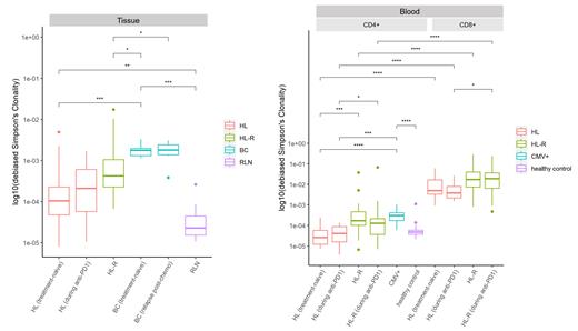
In tissue biopsies, treatment-naïve HL showed a significantly lower dSC compared to treatment-naïve BC and significantly higher dSC compared to RLN from healthy controls. No significant differences could be observed comparing HL with different treatments (during anti-PD1 vs. after chemotherapy) and treatment-naïve HL vs. HL during anti-PD1 treatment. HL and HL-R biopsies showed a significantly lower clonal expansion of Non-Singletons than BC tumors.
In peripheral blood, healthy controls, treatment-naïve HL and HL patients during anti-PD1 treatment showed significantly lower dSC in their CD4+ T-cells compared to CMV+ controls and also compared to HL-R patients. We identified significantly higher dSC in CD8+ T-cells compared to CD4+ T-cells in the blood of HL patients before and during any treatment and in relapsed HL. During anti-PD1 treatment, CD8+ T-cells showed a significantly higher clonal expansion of Non-Singletons than CD4+ T-cells in HL-R and a similar but not significant trend in HL.
All observed differences in dSC were significant for the PoS too, but as expected by their respective definitions with an opposing pattern (i.e. dSC high = PoS low).
Discussion
We did not observe features of intratumoral T-cell expansion in primary HL samples, early during anti-PD1 treatment or at relapse after chemotherapy, suggesting that within the HL tissue, T-cell expansion is hampered. However, patterns of T-cell expansion differed between TME and the peripheral blood, where we observed features of CD8+ T-cell clonal expansion, already prior to any treatment.
In summary, our findings suggest a possible anti-tumor immune response of CD8+ T-cells in the peripheral blood of HL patients, that is not found in the TME.
Disclosures
Reinke:Adaptive Biotechnologies: Other: Q1 2021 immunoSEQ® Young Investigator Award. Borchmann:Amgen: Consultancy, Research Funding; Novartis: Consultancy, Research Funding; Roche: Consultancy, Research Funding; Merck Sharp & Dohme: Consultancy, Research Funding; Bristol-Myers Squibb: Consultancy; Takeda Oncology: Consultancy, Research Funding; MPI: Research Funding. Bröckelmann:BeiGene: Consultancy, Honoraria, Research Funding; Celgene: Other: Travel Grant; BMS: Honoraria, Research Funding; MSD: Honoraria, Research Funding; Stemline: Consultancy, Honoraria; Takeda: Consultancy, Honoraria, Research Funding.


This feature is available to Subscribers Only
Sign In or Create an Account Close Modal